Gel Documentation
Accurate imaging and analysis of nucleic acids and proteins are essential in molecular biology laboratories for research, diagnostics, and quality control. A Gel Documentation System (Gel Doc, Gel Imager, or Gel Image System) is a specialized imaging device for capturing, analyzing, and documenting DNA, RNA, and protein bands separated through gel electrophoresis. These systems are widely used in Tajikistan’s research labs for their ability to enhance fluorescence-based detection of biomolecules.
A high-quality gel documentation system includes a UV transilluminator, a CCD or cooled camera for high-precision imaging, and an integrated darkroom hood to protect users from UV exposure. Advanced Gel Doc systems offer fluorescence and chemiluminescence detection, along with instant printing, Wi-Fi connectivity, and high-resolution image capture for accurate and reproducible analysis.
For laboratories in Tajikistan looking for a reliable and efficient Gel Imaging System, selecting the best model is crucial for genetic research, protein analysis, and Western blot documentation. Whether for academic research, clinical diagnostics, or pharmaceutical applications, investing in the right Gel Documentation System ensures high-quality and reproducible results.
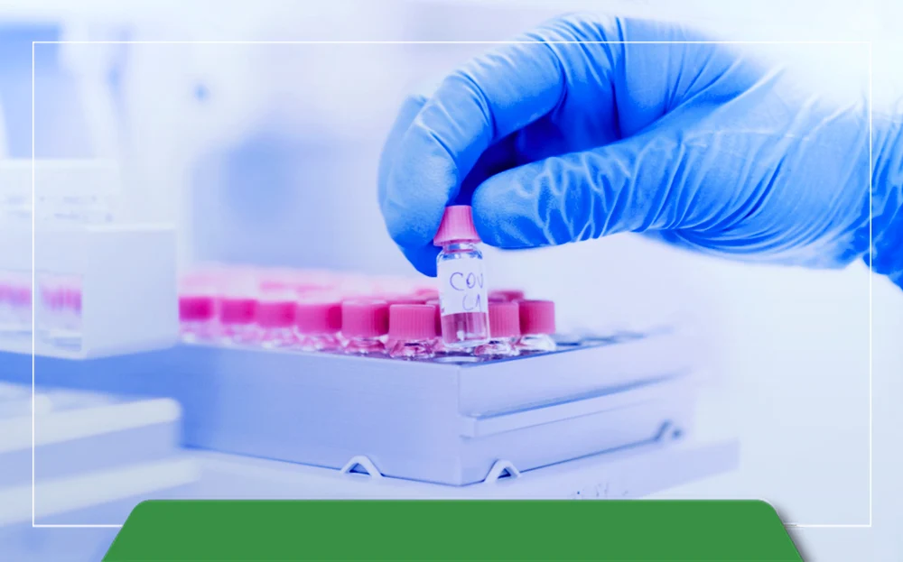
A Gel Documentation System, also known as a Gel Doc or Gel Imaging System, is an essential tool in molecular biology laboratories for capturing and analyzing DNA, RNA, and protein samples. It works by detecting fluorescent signals emitted from nucleic acid or protein samples that have been separated using gel electrophoresis. To visualize these samples, they are typically stained with fluorescent dyes like Ethidium Bromide (EtBr) or SYBR Green, which bind to nucleic acids and emit light when exposed to UV or blue light transilluminators.
At the core of the system is a high-resolution camera, usually a CCD or CMOS sensor, that captures detailed images of the glowing bands. Advanced Gel Documentation Systems use cooled cameras to prevent overheating and enhance sensitivity, allowing for clearer detection of faint bands that might be invisible to the naked eye. Some modern systems even offer chemiluminescence imaging, which is particularly useful for Western blot analysis, helping researchers study proteins and antibody interactions with high precision.
Today’s Gel Documentation Systems come with user-friendly features like touchscreen controls, automatic image enhancement, Wi-Fi connectivity, and instant printing options, making them ideal for efficient lab workflows. In Tajikistan, where laboratories are increasingly investing in modern research tools, choosing the right Gel Documentation System ensures accurate imaging and reliable experimental results.
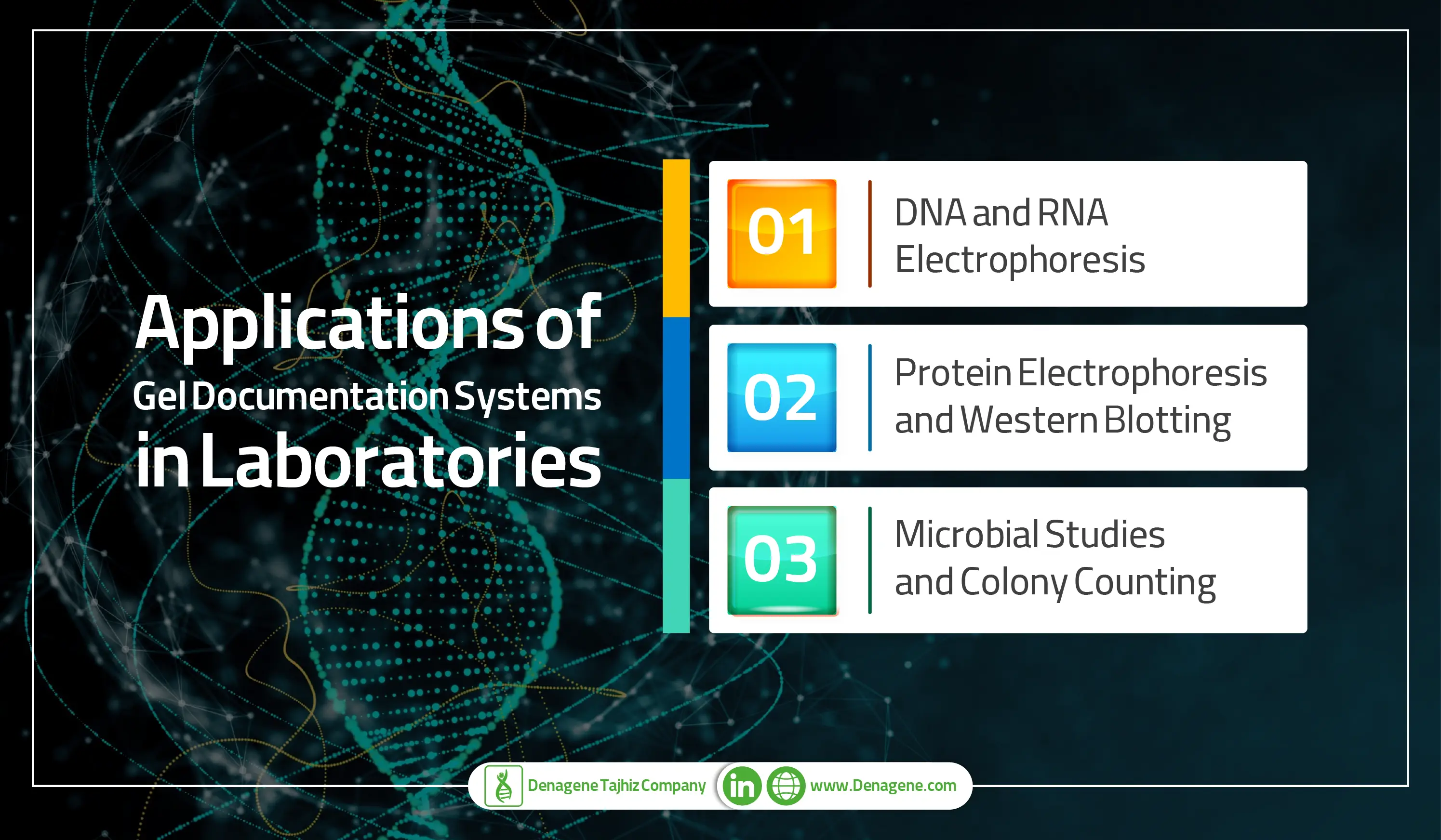
A Gel Documentation System (Gel Imager) is an essential tool in molecular biology, microbiology, and proteomics, allowing researchers to visualize and analyze DNA, RNA, and protein samples with high accuracy. These systems help in various laboratory applications, from electrophoresis analysis to microbial colony counting, making them indispensable in research labs, clinical diagnostics, and genetic studies. Below are the key applications of Gel Documentation Systems in modern laboratories.
One of the primary applications of Gel Documentation Systems is in the analysis of DNA and RNA samples through gel electrophoresis. In this process, nucleic acid fragments are loaded onto an agarose or acrylamide gel and exposed to an electric field, causing negatively charged molecules to migrate towards the positive electrode. Smaller DNA or RNA fragments travel faster than longer ones, enabling researchers to identify specific genetic sequences.
These imaging systems play a crucial role in:
By visualizing DNA bands stained with fluorescent dyes like Ethidium Bromide or SYBR Green, researchers can determine genetic variations, mutations, and molecular weight with precision.
Similar to DNA and RNA analysis, proteins are separated through SDS-PAGE (Sodium Dodecyl Sulfate-Polyacrylamide Gel Electrophoresis) based on molecular weight. Gel Documentation Systems help researchers visualize protein purity, molecular weight, and efficiency of extraction.
Additionally, in Western Blotting, proteins are transferred from SDS-PAGE gels onto membranes for antibody detection. Using a Gel Imager, researchers can analyze:
With advanced chemiluminescence and fluorescence imaging, high-resolution Gel Documentation Systems enhance Western blot analysis for precise protein quantification.
Microbiologists rely on Gel Documentation Systems to analyze microbial diversity and ecology. Additionally, these systems are used for colony counting, determining the number of colony-forming units (CFUs) in petri dishes containing microbial cultures. This application is widely used in:
By automating colony counting and DNA visualization, modern Gel Documentation Systems enhance the speed and accuracy of microbial research.
When selecting a high-quality Gel Documentation System, it is essential to consider performance, durability, and imaging precision. The Gel Documentation System from Denagene Tajhiz is designed with cutting-edge technology to enhance nucleic acid and protein visualization while ensuring environmental sustainability.
For laboratories in Tajikistan looking for the best Gel Documentation System, Denagene Tajhiz’s Gel Doc offers unparalleled imaging accuracy and user-friendly functionality. Learn more about our high-performance Gel Documentation System and how it can enhance your lab’s molecular research.
For pricing, shipping, and order details from Iran to Tajikistan, contact us today!